What is APMPPE?
Apmppe effects young adults between the ages of 20 and 40 who have had a recent bout of viral or flu-like illness? Have you experienced a rapid loss of vision that eventually has affected both eyes?
Have you noticed blotchy scotomas (blind spots) and Photophobia? You may have Acute Posterior Multifocal Placoid Pigment Epitheliopathy.
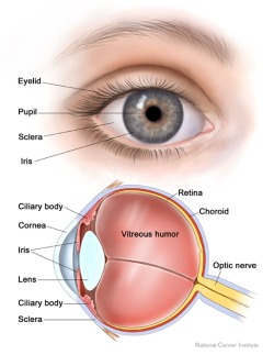
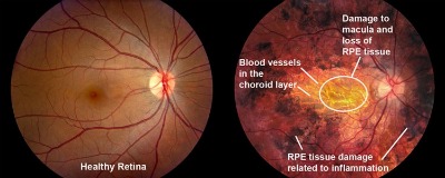
This is an inflammatory eye condition affecting the chorioretinal area of the eye, which includes the Retina and the outermost layer known as the Choroid, which is rich in blood supply.

The cause of the disease is unknown, but is believed to begin with an interruption to the blood supply of the small choroidal arterioles. The deficiency of blood supply that results produces a disruption of the retinal pigment epithelium (RPE).
This is a relatively benign condition that affects both eyes and usually affects otherwise healthy young adults (average age of 25-27 years). These young adults usually have a history of viral illness preceding the onset of visual symptoms. The disease is self-limited.
Less commonly, an association with concurrent disease of the central nervous system (CNS) has been described in the literature.
Among the CNS conditions reported are:
- Meningitis
- Encephalitis
- Vasculitis
- Stroke
- Sinus thrombosis (Sagittal and Cavernous)
What are the Signs and Symptoms of APMPPE?
APMPPE is characterized by multiple yellow-white placoid (flattened) subretinal lesions of the posterior pole. These lesions block out background choroidal fluorescence during the early stages of Fluorescein Angiography (FA). The late stage of FA demonstrates an accumulation of dye in the diseased RPE.
FA, a diagnostic test in which a dye is injected into the arm and the flow of this dye as it enters the eye is studied, shows a choroidal flush which is normally bright as the rich vasculature of the choroid lights up in normal individuals.
This disease involves the RPE, particularly in the Macula area, causing visual loss with subsequent spontaneous resolution. The lesions described above frequently occur in both eyes and in various stages of evolution, typically resolving in weeks to months and leaving residual pigmentation.
If the Macula is affected, vision may be significantly decreased but recovery is the norm, with 80% of those eyes affected achieving a visual acuity of 20/40 or better.

If you have this disease, you may experience any of the following symptoms:
- Rapid loss of vision eventually affecting both eyes.
- Scotomas will present corresponding to the locations of the lesions.
- There may be a relationship with a recent viral illness.
Other early stage symptoms:
Photopsia which is the sensation of seeing lights, sparks, or colors and which is caused by diseases of the Retina or brain
Metamorphopsia/micropsia refers to the distorting of vision (such as straight lines appearing wavy) or objects appearing smaller
Photophobia
In rare cases, redness of the Conjunctiva or Episcleritis.
Late stage symptom is commonly mild visual impairment (20/25 to 20/40). Significant visual loss (20/200) is rare.
How is APMPPE Managed and Treated?
This disease commonly shows rapid healing of the lesions of the Retina over one to two weeks. Symptoms such as vision compromise or visual field defects may persist for up to six months. The vast majority of patients recover to an acuity level between 20/20 to 20/30. Thus, no treatment is used for the retinal findings alone.
If your disease is accompanied by Anterior Uveitis or other eye inflammatory condition, your eye doctor will treat the inflammation appropriately. Your eye doctor should also monitor you for any new blood vessel growth in your choroid as a result of any chronic blood flow deficiency of the choroidal vessels.
If you develop APMPPE and have any signs or symptoms suggestive of a neurological or systemic disorder, make sure your doctor evaluates you carefully for CNS conditions or refers you to a specialist for evaluation for any CNS processes known to be associated with the disease.
Even though APMPPE is generally benign, it can in rare cases be an indicator of serious, possibly life-threatening CNS pathology such as those mentioned above. If your doctor fails to investigate any neurological or body disorder presenting with APMPPE, it can have devastating consequences.


