|
What is a Macula Hole?
Have you experienced a blurring of your central vision? Have you noticed a central blind spot or a gray area in your central vision? You need to go immediately to your eye care provider to make sure your symptoms are not due to a Macula Hole. This is a problem in the central portion of the retina. The severity of the symptoms is dependent on whether the hole is partial or full-thickness. 
For example, partial holes only affect part of the macula layers, resulting in wavy, distorted, and blurred vision. On the other hand, full-thickness holes can cause a complete loss of central vision. Interestingly, this occurs more frequently in women than in men although the reason for the gender difference is unknown. What are the causes?
Although the exact mechanism for the formation of Macula Hole is unknown, some factors have been implicated: In terms of aging, the cause points to the Vitreous. The eye is not hollow on the inside. It contains a gel called the Vitreous. With age, this gel separates into fibrous strands and liquid-like parts as well as shrinks away from the retina. On the macula (the center of the retina), the Vitreous adheres well, and so when the gel shrinks and pulls away from the retina, it can cause a hole. --In the --In order to view the content, you must install the Adobe Flash Player. Please click here to get started.
How is it diagnosed in the eye?
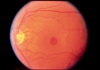
Of course, if you experience a sudden blurring of central vision or a central wavy distortion of your vision, you will want to immediately see an eye care provider. If you have experienced some type of eye trauma, you are in an emergency situation because a number of vision threatening conditions result from trauma. This can be diagnosed during a routine eye exam. Testing vision, Amsler Grid Testing (which tests the central field of vision for waviness, distortion or blind spots), and a dilated fundus examination (DFE) are all performed to evaluate the macula’s health and function. A DFE involves viewing the retina with special high-powered lenses and can reveal the central defect shown in the picture above. Technology has enhanced the ability to confirm retinal findings viewed during a DFE on a histological level. Optical Coherence Tomography (OCT) is a noninvasive imaging technology that provides cross-sectional images of the retina and can help the eye doctor differentiate between partial and full-thickness holes. How is a Macula Hole treated?
An impending hole can spontaneously close, remain stable, or progress to a full-thickness hole. In most cases, surgery is necessary to close the hole and restore useful vision. Surgery often includes vitrectomy, fluid-gas exchange and face down postoperative positioning. Vitrectomy is a procedure in which the gel of the eye is removed gently. This eliminates any traction on the macula. A gas bubble is injected in the eye where the Vitreous was in order to place gentle pressure on the macula and help the hole to seal. In order to help the recovery process after the surgery, you will be required to stay in a face-down position. This period can vary from a few days to three weeks. This enables the bubble to rest on the macula, thereby plugging the hole. The procedure like any other has some potential complications to keep in mind. The most common complication is Cataract. Other complications include: Because Cataract is such a common complication, some surgeons opt to remove the lens prior to or simultaneous to the surgery. |


