|
What is Vitrectomy?
Have you noticed spots in your field of view that will not disappear? Do you have diabetes? Have you sustained a traumatic blow to your eye? Have spots in your field of vision persisted since your injury? You may need to have a surgical procedure known as Vitrectomy. 
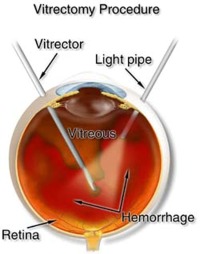
The inside of the eye is not hollow. As you can see from the diagram above, there is a gel that is found behind the crystalline lens and in front of the Retina (the tissue in the back of the eye). This gel is known as the Vitreous. Vitrectomy is a procedure in which the gel of the eye is removed gently. 
Age causes the Vitreous gel to break down. This process results in the jellylike substance getting more fibrous in some areas and more liquid-like in others. When a bright light source (like the sun) shines in your eyes, it may cast a shadow of these clumps and strands of Vitreous visible to you as floaters. Persistent Floaters that do not resolve can result from a number of causes: If the spots in your Vitreous are due to blood, Retinal Detachment, chronic inflammation or signs of cancer, you may need a Vitrectomy. This is used to clear Vitreous opacities or prevent vitreoretinal traction. Vitreoretinal traction is the physical pulling of the Retina by the Vitreous. If you have a Retinal Detachment, the detached tissue, along with any blood and debris, may make it difficult for your Ophthalmologist (preferably Retinal surgeon) to adequately access the back of the eye and repair the detachment. Vitrectomy will help the surgeon to get the necessary access. Suppose you have Diabetic Retinopathy, you may have a Vitrectomy to remove blood from a Vitreous hemorrhage that does not clear. This is also a procedure used to treat Macula Hole because it eliminates any traction on the Macula. A gas bubble is injected in the eye where the Vitreous was in order to place gentle pressure on the macula and help the hole to seal. In order to help the recovery process after the surgery, you will be required to stay in a face-down position. This period can vary from a few days to three weeks. This enables the bubble to rest on the macula, thereby plugging the hole. The procedure like any other has some potential complications to keep in mind. The most common complication is Cataract. Other complications include: Because Cataract is such a common complication, some surgeons opt to remove the lens prior to or simultaneous to the Macula Hole surgery. Surgery can also be performed to treat the complications of Anterior Uveitis. Some researchers noted a positive effect of Vitrectomy on the resolution of chronic Uveitis and recommended the procedure if the inflammation persisted after a few months of oral steroid treatment. This surgery can be utilized to biopsy the Vitreous for diagnostic purposes, be it to rule out infection, inflammation, or cancer. In a nutshell, This surgery is used to treat Macular disorder, Diabetic Retinopathy, Vitreoretinal Traction, and Vitreous Hemorrhage. It can be useful in managing chronic inflammation or in ruling out cancer in the eye, as seen in Retinoblastoma. Floaters due to age are generally benign; however, symptoms of Floaters that persist can indicate other serious eye diseases.
|


